May-Thurner Syndrome Treatment
What is May-Thurner Syndrome, and how does it happen?
When your left iliac vein is trapped between your right iliac artery and the spine, MTS (also known as Iliac Vein Compression Syndrome) occurs. Blood flow is restricted, and pressure and discomfort in the left pelvis and leg increases as a result.
Your left leg is more prone to Deep Vein Thrombosis (DVT) because of this sluggish flow and increased pressure. A DVT is a blood clot that can be quite dangerous. MTS has been linked to the development of Pelvic Congestion Syndrome in women and Varicocele in males.
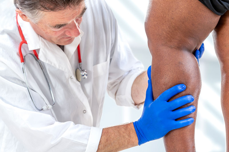
Symptoms of May-Thurner Syndrome
- Pain in legs
- Swelling in legs
- Feeling of heaviness in legs
- Venous claudication, which is leg pain when walking
- Discoloration of the skin
- Ulcers on legs
- Legs with enlargement of the veins
- Pain in the pelvis, back, or hips
What treatment is available for May-Thurner Syndrome?
There are currently no effective surgical procedures to address this problem. Thankfully, at MIVA, we provide a minimally invasive out-patient procedure that is both safe and effective.
We will do a venogram to place a stent in the left iliac vein and keep it open to enable normal blood flow after imaging evaluation and clinic consultation. This therapy has an excellent success rate of 97%.
What is a venogram with intervention and how does it work?
“Veno” comes from the Latin word for vein, and “Gram” is a term that means image. Our doctors use x-ray technology to “see” your veins and assess their functionality to determine the appropriate treatment plan using ultrasound and X-ray guidance. Ultrasound is used to access a vein in either your arm, leg, or neck with a hollow needle after determining the best location. A wire is then pushed through the vein using X-ray assistance.
Following that, there are a variety of treatments available to you, depending on your situation:
- Angioplasty is a type of procedure used to unclog blockages or narrow veins. A wire may be placed in the vein and inflated with specialized balloons to open clogged areas or constricted veins.
- A stent, which is tubular metal scaffolding, may be placed in the vein to keep it open if the vein does not maintain its own opening.
- Embolization can be used in the case of a blocked vein. We can use coils or plugs to seal it off.
- Thrombolysis: If there is a substantial quantity of clot (a condition known as Deep Vein Thrombosis, or DVT) in the arm, leg, stomach, etc., a special catheter can be inserted into the clot and deliver medication that dissolves the clot on touch. This may be done in a single day if the patient is placed in an ICU.
Internal examination of the veins with Intravascular ultrasound (IVUS) is possible in specific situations.
Procedures such as those are done as outpatient operations at MIVA or at a local hospital. The treatment might need general anesthesia in some situations, but it frequently can be completed with moderate sedation. Recovery time is fast since there are no incisions. The major barrier is that you will not be able to perform significant lifting (defined as > 15 lbs.) for five days after the treatment. Blood thinners may be required on following the therapy.
FAQ's
What illnesses can a venogram help with?
The list is lengthy, but the following are some of the more typical diseases treated:
- Pelvic Congestion Syndrome
- Acute and Chronic DVT
- Superior Vena Cava (SVC) Syndrome
- Varicocele
- May-Thurner Syndrome